Viruses, Nutritional Immuno-Modulators and Metabolic Types
The immune system is a complex network consisting of cells, tissues and organs and their coordination is required to protect the body from infectious pathogens or viruses and non- infectious foreign substances. It is essential for the immune system to be at its optimum for resistance to invading pathogens and for the development of immunity. Optimisation of the immune system depends upon many factors, not the least of which includes our nutritional status, but also the psycho-neuro-endocrine system.
Thank you for reading this post, don't forget to subscribe!As Viewed through Hair Tissue Mineral (HTMA) Patterns – By David L. Watts, Ph.D., Director of Research TEI Laboratories & Head Research Consultant InterClinical Laboratories.
Branches of the Immune System
The immune system responds as a whole normally, but in order to simplify we can view the immune system being made up of two components or branches. They are the cellular and humoral branches, also called innate and adaptive immunity. Typically, the first response against microbes is initiated by the cellular immune response, followed by the adaptive or humoral response. Completion of this two- step coordinated process ultimately confers adaptive or specific immunity to ward off invading microbes as well as provide memory to the cells in order to respond to the same microbe in the future due to the production of specific antibodies, thereby conferring immunity to subsequent exposure. Rather than discuss the primary and secondary organs and cells of the immune system they can be found here;
https://interclinical.com.au/wp-content/uploads/2020/03/1994-March-April-The-Immune-System-and-Hair-Tissue-Mineral-Patterns.pdf
Or you can review other immunology, biochemistry or physiology texts. To simplify further, I will use the terms cellular immune response (CIR) and humoral immune response (HIR). When we view the CIR and HIR in relationship to the HTMA mineral patterns, we have discerned that the CIR is associated with a parasympathetic and the HIR is associated with a sympathetic HTMA mineral pattern. This is important to note as the immune system is affected by the neuro- endocrine system and vice-versa. Further, the CIR generally speaking, is initiated by the thymus and the HIR is adrenal mediated. Of course, other organs and endocrines are involved in both responses.
Immune Imbalance
When the immune system is not compromised and has dealt with a threat, the systems return to a homeostatic balance. I rather use the term rheostatic balance as cells and biochemical defense systems in body are constantly alert, on guard and ready to mount a defense toward an invader, similar to a standing army. However, it is well known that a branch of the immune system can become overactive and become detrimental to the host. This is due to the potential antagonism between the CIR and HIR immune response causing autoimmune conditions. Further discussions can be found in the following link;
https://interclinical.com.au/wp-content/uploads/2020/03/2002-Jan-Feb-Autoimmune-and-Women.pdf
In summary, the CIR (thymus-parasympathetic) can become dominant suppressing the HIR (adrenal-sympathetic). Conversely, the HIR (adrenal-sympathetic) can become dominant and suppress the CIR (thymus-parasympathetic). Other researchers have found this to be the case. Harrison, found an inverse relationship between humoral and cellular mediated immunity in subjects at risk of insulin dependent diabetes.
It is speculated that in an autoimmune disease, such as insulin dependent diabetes, the pancreatic beta cells are destroyed or impaired by the T-cells, which is associated with cell-mediated immunity. It is interesting that HTMA patterns of patients with adult onset diabetes, are commonly associated with the slow metabolic mineral pattern, or increased cellular immune activity. Other studies have reported finding an imbalance between the cellular and humoral response in conditions such as lupus, rheumatoid arthritis, Sjogren’s Syndrome, Crohn’s disease and celiac disease.
In other words, it is very evident that the cellular immune response can over-power the humoral immune response and vice-versa. The following statement by Katz illustrates this point very well, “..it is becoming increasingly well documented that, in some animal models and in humans, the humoral immune system can act to the detriment of the host and indeed, block the activity of the cellular immune system in its attempt to destroy neoplastic cells.”
https://interclinical.com.au/wp-content/uploads/2020/03/1994-March-April-The-Immune-System-and-Hair-Tissue-Mineral-Patterns.pdf
When discussing or treating autoimmune conditions, one must consider which branch of the immune system to support rather than generally treating the immune system as a whole.
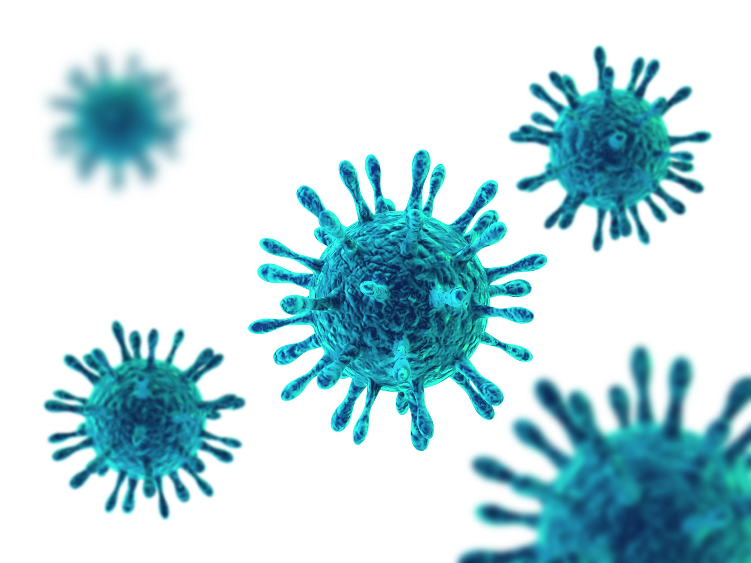
Viruses and the Immune System
Viruses activate the CIR, which I mentioned previously is also a thymus response in tandem with a parasympathetic response. Bacteria however tend to activate the HIR in conjunction with an adrenal and sympathetic response. As viral infections are more common than bacterial infections I will limit this discussion to viral conditions.
HTMA Patterns and CIR
HTMA studies have shown distinct mineral patterns associated with an elevated CIR and autoimmune conditions associated with excessive CIR. As mentioned previously, the CIR is associated with a parasympathetic mineral pattern and it can be surmised that a parasympathetic mineral pattern may predispose the body to viral susceptibility due to a number of factors.
Elevated Tissue Calcium and Viral Activity
One factor that increases viral susceptibility is elevated tissue concentrations of calcium. Viruses use the calcium signals to enter cells and to create an ideal environment for their replication. (Cell Calcium, 2009)
“The exact mechanism behind viral latency or viral replication is not totally known. However, a group of researchers explored this phenomenon in relation to the Epstein Barr virus, and their findings were reported in SCIENCE in 1986. Lymphoid cells were infected with the Epstein Barr genome. The Epstein Barr virus would either replicate slowly or remain dormant. Dormancy of the virus could be overcome a number of ways such as super-imposing a super virus or by introducing tumour promoting agents. The tumour promoting agents that activated the Epstein Barr virus also activated a cellular enzyme protein kinase C that is calcium dependent; therefore, they studied the effect of increased cellular calcium concentration on the lymphatic cells. They found that calcium modulation was of primary importance in activating the Epstein Barr genome. Other studies have been reported that implicate calcium in regulating the infection of human B-lymphocytes by Epstein Barr virus. When calcium entry into the cells was blocked, the Epstein Barr virus transformation was inhibited.”
https://interclinical.com.au/wp-content/uploads/2020/03/1989-Nov-Dec-Calcium-and-Virus-Activation.pdf
The Journal of Virology (2010) published findings that the West Nile (WNV) virus infections lead to a rapid and sustained calcium influx into the cells resulting in extending the survival of the WNV within cells. They found that reducing the influx of calcium into cells by various methods during an early infection decreased viral yield.
Calcium Antagonism and Viral Inhibition
It is apparent that reducing cellular calcium influx would also help to reduce cellular viral influx and replication. Interestingly, hydroxychloroquine, an anti-malaria drug can contribute to anti-viral activity by reducing cellular calcium influx. (Chloroquine Inhibits Ca2+ Signalling in Murine CD4+ Thymocytes. https://www.researchgate.net/journal/1421-9778_Cellular_Physiology_and_Biochemistry)
Also interesting is that elevated calcium concentrations are found in those with sickle cell disease, a condition associated with malaria. (Prog Clin Biol Res.) (Sci Rep. 2017) There are many drugs designated as calcium channel blockers which one can wonder if they may impact viral replication by their action. Apparently, calcium channel blockers such as verapamil increases the concentrations of chloroquine in the cell. Of course there are natural nutritional calcium blockers available such as phosphorus, magnesium, and Vitamin A.
Latent Epstein-Barr Virus (EBV) is present in approximately ninety percent of humans worldwide and is frequently established following a viral episode such as mononucleosis. EBV is a member of the herpes virus family. Usually dormant, chronic EBV activation can lead to lymphoma, and other health conditions. Recent investigations have found that in individuals who have a genetic defect in the magnesium transporter 1 have high levels of EBV.
Conversely, individuals with chronic EBV infections had low amounts of basal free magnesium levels in their cells, even though bound levels of magnesium were found to be normal. Ninety-five percent of the magnesium present in the cell is bound and the rest is in a free form. A reduction in free cellular magnesium concentration is associated with a defect in the expression of the natural killer activating receptor in natural killer cells, CD8 T cells and reduces the cytotoxic response against EBV. With magnesium supplementation, a restoration of the concentration of free cellular magnesium results in reduced EBV infected cells. Therefore, magnesium homeostasis is important for antiviral and antitumour immunity. (Science. 2013)
Copper Excess and Pro-Viral Activity
We consider copper as a pro-viral agent. It has an antagonistic effect on the mineral zinc which is known to have anti-viral properties. We often see elevated HTMA copper in individuals who have had or who are suffering from viral episodes, including mononucleosis, AIDS, EBV, CMV, etc. Copper also acts synergistically with calcium and enhances increased tissue calcium accumulation and thereby, predispose the cell to viral replication. (Virol J. 2017) I realise there are a lot of statements on the internet promoting copper as an anti-viral agent which I find a bit questionable.
Copper has long been known for its antimicrobial properties particularly toward bacteria. Bacteria such as staph, MRSA and C. difficile, etc. were destroyed almost immediately when exposed to copper surfaces with almost 100 percent within a couple of hours which is why many hospitals use copper coated surfaces and bed rails which has reduced hospital-acquired staph infections tremendously. While some viruses are known to take up to 4 hours to degrade on copper surfaces. It is possible I suppose that the effect of copper on viruses could be due to differences between RNA and DNA viruses.
Anti-Viral Properties of Zinc, Molybdenum and Vitamin C
Zinc is also well known for its anti-viral properties. Zinc is also antagonistic to the mineral copper. Perhaps the reason that chloroquine has some anti-viral effects is due to its ionophore action. Chloroquine enhances the transport of zinc across cell membranes and into cells. Zinc is a strong inhibitor of RNA viruses by inhibiting RNA polymerase. Pneumonia is a major cause of death in children less than five years of age. A double-blind placebo-controlled clinical trial of 270 children found that zinc supplementation in the amount of 20 milligrams per day resulted in accelerated recovery from pneumonia.
The mineral zinc also helped to reduce antimicrobial resistance and decreased multiple antibiotic exposure as well as reduce complications and deaths. (Brooks, et al. Lancet, 363, 2004) Low zinc level during a viral infection is often associated with a loss of taste and smell, and is common in most severe viral infections. White spots showing up in the fingernails following a viral infection are also an indication of zinc deficiency.
Vitamin C and molybdenum are also known to be anti-viral due to their copper antagonism. Both zinc and vitamin C have been reported to be effective in treatment of those who have other viruses. A number of studies are being carried out in the U.S. and abroad on the therapeutic effectiveness of zinc and vitamin C in patients infected by a virus.
Anti-Viral Properties of Niacin and Vitamin A
We have found that HIV is associated with elevated tissue concentrations of copper in a number of studies we have performed at TEI. As stated previously, we categorize excess copper as being pro-viral. Excess copper increases the oxidation of vitamin C and can contribute to pellagra. It has been found that individuals who have contracted HIV virus progressing to AIDS and ARC, develop a pellagra-like condition. Tryptophan levels are extremely low along with niacin contributing to NAD depletion. Dietary niacin therapy led to an increase in tryptophan levels in those affected. Normally tryptophan is converted to niacin, but providing tryptophan would potentially increase concentrations of neurotoxic intermediates. (Lancet. 2003) (Nutrition. 2001)
Vitamin A is synergistic to zinc and also lowers tissue calcium and is therefore, considered to be anti-viral. Vitamin A is known to combat respiratory tract viral infections that contribute to pneumonia. One mechanism for vitamin A’s effectiveness is that it can contribute to down regulation of IL-6 and reduce hyperactive T cell response to viruses and reduce the hyperactive cytokine storm. (Penkert,R, et.al. 2017) (Liang, Y, et al. 2013) Vitamin A is essential for healthy mucous membranes, the first defensive barrier to microorganisms.
Cytokine Storm and Hyperactivity of the CIR
Cytokine production is a normal consequence of viral infections. However, when the cellular immune response becomes over active, a cytokine storm can develop and be detrimental to the host. On the basis of research into the immune system, Notkins and colleagues made the following statement, “…there is growing evidence that cells are damaged not directly by replicating viruses but by a specific immune response that produces the symptoms of the disease.”
The authors site Clemens von Pirquet, an Austrian paediatrician who more than 60 years previously, speculated that the immune response may be responsible for injury to tissues. Wallace P. Rowe, a virus researcher at the National Institutes of Health, later confirmed von Pirquet’s suspicions. His experiments involved the study of the lymphocyte choriomeningitis (LCM) virus. When introduced into lab animals the virus spread rapidly throughout the body. In six days the animals began to show an immune response to the virus, developed meningitis and died. To determine the immune systems involvement, Dr. Rowe performed another experiment, in which he exposed one group of animals to radiation, as radiation is known to suppress the immune response. Whereas, in the control group, the immune system was not suppressed. After the LCM virus was introduced into the animals, the virus spread throughout the bodies of both groups.
The control animals developed meningitis and died, as in previous experiments. However, the immune suppressed group did not develop meningitis. Rowe, therefore found that the meningitis was caused not by the virus itself, but by the body’s immune response to the virus.
https://interclinical.com.au/wp-content/uploads/2020/03/1994-March-April-The-Immune-System-and-Hair-Tissue-Mineral-Patterns.pdf
In order to control the cytokine storm seen in some viral cases, immune-suppressants are used effectively, such as steroids, and other immuno-suppressing drugs.
Adverse Effects of Cellular Immune Stimulation and HIV
Researchers have been working on vaccines that would produce a cellular immune response and not a humoral immune response in relation to HIV-specific cytotoxic T-cell response. In doing so, they hoped that the vaccine may lower viral set point and slow disease progression in those who acquired HIV. However, in the trial vaccines neither prevented infection or lowered viral set point. In fact, the recipients of the vaccine appeared to be at higher risk for HIV acquisition and had a two-fold increase in the incidence of infection. As a result, the international trials were stopped. (Del Rio, C. 2007) These results support our findings at Trace Elements through HTMA studies that AIDS is associated with an autoimmune or overactive cellular immune response. Instead of supporting the thymus (cellular immune response), focus should be on suppressing it and or supporting humoral immunity which is typically suppressed in ARC and AIDS patients.
Trace Elements and the Immune System
Lukac, et al reported the importance of trace elements on the immune system. They play an important role in physiological processes that are crucial for normal functioning. The authors also state that deficiencies of trace elements are often found with infectious disease and can therefore, influence susceptibility, course and outcome of a number of viral infections. Further, “Some trace elements inhibit viral replication in the host cells and therefore have antiviral activity. Many trace elements act as antioxidants or are able not only to regulate the host immune response but also to alter viral genome.” (Lukac, 2007)
It is known that nutritional deficiencies exist in the early stages of human immunodeficiency virus (HIV) infection. Botswana, has one of the highest rates of HIV infection in the world and early treatment in the population with antiretroviral therapy (ART) can be slow and difficult. A trial using multivitamins was conducted involving over eight- hundred HIV patients in the early stages of HIV and who were not on ART. The vitamins included B complex along with vitamins C, E and selenium. The study was conducted over a two year period and found that nutritional supplementation was safe and significantly reduced the risk of a decline in immunity as well as morbidity among those treated compared to a placebo group. (Baum, MK, et al. 2013.)
Viral Complications and Metabolic Types Based Upon HTMA Patterns
Viruses are responsible for most infections in industrialized populations. From HTMA studies that we have performed around the world (over one million) we have found that the majority of individuals have parasympathetic dominant mineral patterns (approximately 70% average) making them more susceptible to complications of viral infections. This mineral pattern is associated with dominant cellular immune activity. The following chart shows the percentage of metabolic types in different countries based upon HTMA patterns we have performed.
Interestingly, it appears that in countries with a higher population of Parasympathetic dominance (CIR), death rates are higher compared to countries with a lower percentage of Parasympathetic dominance (CIR). As of this date, we can see Norway appears to have the lowest percentage of Parasympathetic metabolic types and has fewer deaths per confirmed case of severe viral infection compared to countries such as England and Italy. However, the HTMA metabolic patterns do not take into consideration the age of the population and those other factors that may contribute to vulnerability to the virus.
Although specific nutrients or drugs would not prevent anyone contracting a virus we can surmise that specific nutrients may aid in reducing complications of viral infections. Anti-viral nutrients in individuals with a dominant CIR include; magnesium, phosphorus, zinc, molybdenum, selenium, vitamin C, vitamins A, B1, B3 and E. Synergists to these would also include; vitamin B6, and amino acids such as methionine. Nutrients or products that would further stimulate the CIR such as; calcium, copper, vitamin D, thymus, Co-Q10, colostrum, etc, would be contraindicated, as these nutrients enhance increased cellular calcium concentrations.
For sympathetic types having an elevated HIR (approximately 30% of population) then calcium, magnesium, vitamins D, B2, B1, thymus, colostrum and co-Q10 would be indicated.
We should also remember that the endocrine system is also involved in immune function and balance. A disturbance in the HPA and gonadal axis can impact and produce a compromised immune response which should also be addressed. Some hormones can be categorized as anti-viral or pro-viral. Reports of individuals with viral infections have found that progesterone may help in preventing complications as it appears to help reduce inflammation. Progesterone as well testosterone is associated with the mineral zinc and along with zinc lowers copper as well as estrogen. Hormones that increase parasympathetic activity and CIR dominance could be considered pro-viral and include; Estrogen, Insulin and Parathyroid Hormone. Anti-Viral hormones, those that raise sympathetic activity and HIR activity include; Progesterone, Testosterone, and Thyroid. Further discussion can be found at; https://interclinical.com.au/wp-content/uploads/2020/03/2002-Jan-Feb-Autoimmune-and-Women.pdf
Conclusion
HTMA patterns provide us information regarding mineral relationships. As Dr. Schroder stated in his book Trace Elements and Man, “minerals are the sparkplugs of life.” However minerals also have a double edged sword effect, in that even though a low level can produce risks such as compromising the immune system, excess levels and or disturbance in their balance can also compromise the immune system.
A Word About Vitamin D
Vitamin D is being highly promoted as a major nutrient in combating the effects of some viral infections. However, its role remains uncertain. For vitamin D to be effective it must be used with the proper metabolic type and immune response. For sympathetic types who have an elevated or dominant HIR, vitamin D would be most helpful. In such cases, vitamin D would help to stimulate the CIR and reduce the cytokine inflammation due to excessive HIR. Vitamin D enhances the thymic response and reduces the hyperactive adrenal response.
Vitamin D is contraindicated for individuals who are parasympathetic dominant due to their overactive CIR or excessive thymus activity. Vitamin D is of course a regulator of calcium and phosphorus but is also related to parathyroid hormone (PTH) and insulin. Vitamin D, PTH and insulin, along with excess estrogen can raise tissue calcium concentrations and therefore, allow viral proliferation. Vitamin A on the other hand, would be beneficial in the parasympathetic pattern (CIR) as vitamin A would help to reduce the excessive thymic and CIR reaction as well as lower calcium.
REFERENCES
Cell Calcium, 2009 Viral Calciomics: Interplay between Ca2+ and virus
The Journal of Virology 2010 Virus-induced Ca2+ influx extends survival in West Nile virus infected cells
Prog Clin Biol Res. 1978;20:105-22. Calcium exchange and calcium-related effects in normal and sickle cell anemia erythrocytes. Cameron BF, Smariga P
Sci Rep. 2017 May 15;7(1):1892. doi: 10.1038/s41598-017-01836-8. Pro-inflammatory Ca++-activated K+ channels are inhibited by hydroxychloroquine. Eugenia Schroeder M, et al.
MG Regulates Cytotoxic Functions of NK and CD8 T Cells in Chronic EBV Infection Through NKG2D. Chaigne-Delalande, B, et al. Science, 341, 6142, 2013.
Virol J. 2017; 14: 11. Published online 2017 Jan 23. doi: 10.1186/s12985-016-0671-7 PMCID: PMC5259989 PMID: 28115001 Host Cell Copper Transporters CTR1 and ATP7A are important for Influenza A virus replication.
Brooks, et al. Zinc for severe pneumonia in very young children: Double-Blind Placebo-Controlled Trial. Lancet, 363, 2004.
(Lancet Infect Dis. 2003 Oct; 3(10):644-52. Tryptophan depletion and HIV infection: a metabolic link to pathogenesis. Murray MF.
Nutrition. 2001 Jul-Aug; 17 (7-8):654-6. Increased plasma tryptophan in HIV-infected patients treated with pharmacologic doses of nicotinamide.
Penkert, R, et.al. Vitamin A differentially regulates cytokine expression in respiratory epithelial and macrophage cell lines. Cytokine, Mar. 2017.
Liang, Y, et al. Retinoic Acid Modulates Hyperactive T Cell Responses and Protects Vitamin A-Deficient Mice against persistent Lymphocytic Choriomeningitis Virus Infections. J. Immun. Apr. 2013
Del Rio, C. Aids Clin. Care Nov. 2007.) (Lukac, n, Massanyi, P. Effects of Trace Elements on the Immune System. Epidemiol. Mikrobiol. Immunol. 56,1,2007.
Effect of Micronutrient Supplementation on Disease Progression in Asymptomatic, Antiretroviral-Naïve, HIV-Infected Adults in Botswana. A Randomized Clinical Trial. Baum, MK, et al. JAMA 310,20, 2013.
InterClinical Laboratories Copyright 2020